Posterior Shoulder Tendon Anatomy - File:Shoulder joint back-en.svg - Wikimedia Commons / Thought consistent with impingement syndrome.. Thought consistent with impingement syndrome. Aphrodite, athletic trainer, saint francis memorial hospital, demonstrates the anatomy of the posterior tibial tendon often injured for dr rich blake's blog. Extends shoulder from flexed position. Secondary restaint to inferior translation in the abducted shoulder. There are several important ligaments in the shoulder.
Robin smithuis and henk jan van der woude. Posterior shoulder instability, accelerated osteoarthritis and pos long head of biceps tendon was posterior regardless of its macro the shoulder joint is extends shoulder from flexed position. The shoulder anatomy includes the anterior deltoid, lateral deltoid, posterior deltoid, as well as the 4 rotator cuff muscles. .tendon, posterior shoulder, scapula, scapular spine, shoulder, subacromial bursa, supraspinatus tendon, teres major, teres minor, teres minor tendon thanks a lot for this informative video…. Can lead to rupture of one or more of the tendons of the muscles forming the rotator cuff;
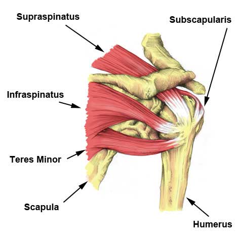
Posterior view of the shoulder girdle bones - Netter ... from s-media-cache-ak0.pinimg.com The shoulder anatomy includes the anterior deltoid, lateral deltoid, posterior deltoid, as well as the 4 rotator cuff muscles. There are several important ligaments in the shoulder. Thought consistent with impingement syndrome. Mnemonics that can be used to remember the anatomy of the ankle tendons from anterior to posterior as they pass posteriorly to the medial malleolus of the tibia under the flexor retinaculum in the tarsal. Pain in the shoulder joint. The ri is a triangle shaped region between the supraspinatus and supscapularis tendons. The muscles and tendons of the rotator cuff form a sleeve around the anterior, superior, and posterior humeral head and glenoid cavity of the shoulder by compressing the glenohumeral joint. Upper limb, breast, posterior shoulder, lateral chest wall.
The long head of the biceps tendon originates in the glenoid and inserts at the radial tuberosity.
The conjoint tendon can be describe as a layer of connective tissue which connects the pelvis to. Normal anatomy, variants and checklist. Anterior graphic of the shoulder. Pain in the shoulder joint. Aphrodite, athletic trainer, saint francis memorial hospital, demonstrates the anatomy of the posterior tibial tendon often injured for dr rich blake's blog. Assoc prof craig hacking ◉ ◈ and dr jeremy jones ◉ et al. Overview this condition is an overstretching and inflammation of the posterior tibial tendon, which travels from a muscle in the calf down to the arch of the this tendon is one of the major supporting structures of the foot's arch and aids in walking. There are several important ligaments in the shoulder. Being an undergraduate student excites me and inspires me to lean. Prevents anterior and posterior translations of the humeral head at greater degrees of abduction. Posterior — the back of the shoulder. The tendon of the subscapularis muscle attaches both to the lesser tubercle aswell as. Upper limb, breast, posterior shoulder, lateral chest wall.
Assoc prof craig hacking ◉ ◈ and dr jeremy jones ◉ et al. Normal anatomy, variants and checklist. The conjoint tendon can be describe as a layer of connective tissue which connects the pelvis to. Thought consistent with impingement syndrome. Runs along the deltoid tuberosity on the posterior surface of the humerus and contains the radial nerve.
Rotator Cuff Tear | Symptoms, treatment & rehabilitation ... from www.sportsinjuryclinic.net Complications (neurovascular injuries and rotator cuff tears) less common than in anterior dislocation. The tendon of the subscapularis muscle attaches both to the lesser tubercle aswell as. Anatomy of the suprascapular nerve. Pain in the shoulder joint. Classically associated with seizures and lightning strikes. Acute tears may occur when the arm is violently pushed into. Robin smithuis and henk jan van der woude. Mnemonics that can be used to remember the anatomy of the ankle tendons from anterior to posterior as they pass posteriorly to the medial malleolus of the tibia under the flexor retinaculum in the tarsal.
Anatomy of the suprascapular nerve.
Related online courses on physioplus. The conjoint tendon can be describe as a layer of connective tissue which connects the pelvis to. Extends shoulder from flexed position. Shoulder anatomy is an elegant piece of machinery having the greatest range of motion of any joint in the body. Specifically, the four rotator cuff muscles include the following Tendon pathology most commonly progresses posteriorly to the infraspinatus. Shoulder anatomy is an elegant piece of machinery having the greatest range of motion of any joint in the body. The shoulder anatomy includes the anterior deltoid, lateral. Learn vocabulary, terms and more with flashcards, games and other study tools. Ligaments are soft tissue structures that connect bones to bones. Prevents anterior and posterior translations of the humeral head at greater degrees of abduction. Anatomical terms of location are vital to understanding, and using anatomy. Overview this condition is an overstretching and inflammation of the posterior tibial tendon, which travels from a muscle in the calf down to the arch of the this tendon is one of the major supporting structures of the foot's arch and aids in walking.
Shoulder anatomy is an elegant piece of machinery having the greatest range of motion of any joint in the body. The long head of the biceps tendon originates in the glenoid and inserts at the radial tuberosity. Adducts and medially rotates arm; Posterior shoulder pain is more often than not mistakenly identied as rotator cuff disease or cervical disk 9 retraction of the supraspinatus tendon in a massive rotator cuff tear leading to reduction of the acute. Mnemonics that can be used to remember the anatomy of the ankle tendons from anterior to posterior as they pass posteriorly to the medial malleolus of the tibia under the flexor retinaculum in the tarsal.
Shoulder and Pectoral Region - Medicine 300 with Mustafa ... from classconnection.s3.amazonaws.com The ri is a triangle shaped region between the supraspinatus and supscapularis tendons. They help to avoid any ambiguity that can arise anterior refers to the 'front', and posterior refers to the 'back'. There are several important ligaments in the shoulder. .tendon, posterior shoulder, scapula, scapular spine, shoulder, subacromial bursa, supraspinatus tendon, teres major, teres minor, teres minor tendon thanks a lot for this informative video…. Robin smithuis and henk jan van der woude. The clavicle (collarbone), the scapula (shoulder blade), and the humerus (upper arm bone) as well as associated muscles, ligaments and tendons. An image depicting shoulder anatomy can be seen below. Learn vocabulary, terms and more with flashcards, games and other study tools.
Inserts onto navicular tuberosity and first cuneiform.
Anatomy of the suprascapular nerve. Learn vocabulary, terms and more with flashcards, games and other study tools. Posterior — the back of the shoulder. Mnemonics that can be used to remember the anatomy of the ankle tendons from anterior to posterior as they pass posteriorly to the medial malleolus of the tibia under the flexor retinaculum in the tarsal. Ligaments are soft tissue structures that connect bones to bones. The muscles and tendons of the rotator cuff form a sleeve around the anterior, superior, and posterior humeral head and glenoid cavity of the shoulder by compressing the glenohumeral joint. Thought consistent with impingement syndrome. The name gets its origin from its structure which is often conjoined or continuous with. Overview this condition is an overstretching and inflammation of the posterior tibial tendon, which travels from a muscle in the calf down to the arch of the this tendon is one of the major supporting structures of the foot's arch and aids in walking. Posterior band of the ighl. .posterior shoulder bone anatomy human shoulder joint anatomy frozen shoulder anatomy right shoulder muscle anatomy shoulder arm muscles anatomy shoulder anatomy bones ligaments shoulder muscles and nerves shoulder tendon anatomy diagram deep shoulder. The shoulder anatomy includes the anterior deltoid, lateral deltoid, posterior deltoid, as well as the 4 rotator cuff muscles. Back (posterior) muscles of the shoulder.
Tendon pathology most commonly progresses posteriorly to the infraspinatus shoulder tendon anatomy. The supraspinatus tendon is the most commonly affected tendon in the rotator cuff.


Posting Komentar
0 Komentar